Results and discussion
Absorption and fluorescence excitation spectra and quantum yields for the eximer formation
The broad feature around 158 nm is assigned as the first allowed transition 10σg→7σu. Several series of sharp bands in the shorter wavelength region than 160 nm are Rydberg transitions from the 5πu 3/2, 5πu 1/2 and 10πg orbitals whose band positions converge to the first (12.42 eV), second (12.89 eV), and third ionization energies (13.65 eV), respectively. The band positions observed from the absorption and fluorescence excitation spectra are listed in the next page along with their assignments.
It is noted that the fluorescence quantum yields (= quantum yields for excimer formation) are high in the entire wavelength region observed and the quantum yield drastically decreases for Rydberg bands. In other words, the quantum yields are high in the region of weak absorbance which may be called background absorption region. The background absorption may be assigned to the transitions to intravalence excited states.
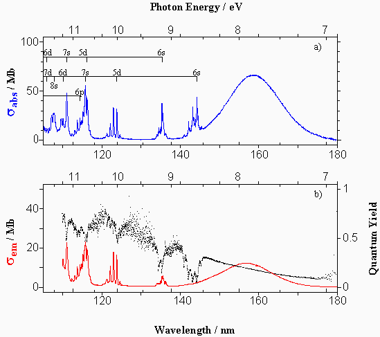
a) Absorption cross section (line), σabs, of XeF2 plotted against the wavelength (bottom abscissa), λex, and photon energy (top abscissa), Eex, of the exciting light. b) Photoemission cross section (line), σem, and the quantum yield (dots) for the total XeF* fragment formation in photodissociative excitation of the XeF2 plotted against λex (bottom abscissa) and Eex (top abscissa). The spectral resolution of the exciting light was Δλex = 0.1 nm.
Fluorescence anisotropy, R
The R value strongly depends on the type of state to which the XeF2 molecule is excited. The present results of fluorescence anisotropy measurements support that the 10σg→7σu, and 10σg→6pσR transitions are parallel and the 5πu→nsR or ndσR transitions are perpendicular to the molecular axis.
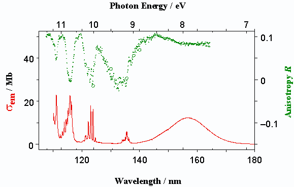
Fluorescence anisotropy (dots), R, obtained for total XeF* emission, whose ordinate is indicated on the right against λex (bottom abscissa) and Eex (top abscissa) along with the photoemission cross section (solid line), σem, whose ordinate is indicated on the left. The spectral resolutions of the exciting light for the anisotropy and fluorescence excitation spectra were 0.50 and 0.10 nm, respectively. The absolute R values (o) determined using the PEM-MCS system are also plotted for comparison.
Dispersed fluorescence spectra
The B−X and D−X bands are observed near λobs = 350 and 260 nm, respectively. The C−A emission was observed around λobs = 460 nm but too weak to be easily identified. It would be interesting to note that, when XeF2 is excited at λex = 135.5 nm and 154.0 nm, no conspicuous D−X bands are identified near λobs = 260 nm in the dispersed fluorescence spectra. As to the B−X bands, it is noted that the higher is the excitation energy, the broader the vibrational distribution becomes, which is inferred from the larger contributions of bound-free bands.
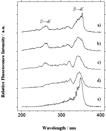
Dispersed fluorescence spectra of XeF* formed at exciting light wavelength, λex, of a) 111.1, b) 115.8, c) 123.8, d) 135.5 and e) 154.0 nm in the observed fluorescence wavelength, λobs, region of 200 ~ 400 nm with a spectral resolution of Δλobs = 1.2 nm. The resolution of the exciting light was Δλex = 1.0 nm.